Diabetic Retinopathy
What is Diabetic Retinopathy?
Diabetes is a disease that affects blood vessels throughout the body, particularly vessels in the kidneys and eyes. When the blood vessels in the eyes are affected, this is called diabetic retinopathy.
The retina is in the back of the eye. It detects visual images and transmits them to the brain. Major blood vessels lie on the front portion of the retina. When these blood vessels are damaged due to diabetes, they may leak fluid or blood and grow scar tissue. This leakage affects the ability of the retina to detect and transmit images.
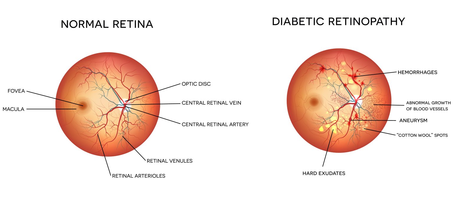
During the early stages of diabetic retinopathy, vision is typically not affected. However, when retinopathy becomes advanced, new blood vessels grow in the retina. These new vessels are the body’s attempt to overcome and replace the vessels that have been damaged by diabetes. However, these new vessels are not normal. They may bleed and cause the vision to become hazy, occasionally resulting in a complete loss of vision. The growth of abnormal blood vessels on the iris of the eye can lead to glaucoma. Diabetic retinopathy can also cause your body to form cataracts.
The new vessels also may damage the retina by forming scar tissue and pulling the retina away from its proper location. This is called retinal detachment and can lead to blindness if left untreated.
Causes of diabetic retinopathy
Diabetes: Everyone who has diabetes is at risk for developing diabetic retinopathy, but not everyone develops it. Changes in blood sugar levels increase the risk. Generally, diabetics don’t develop diabetic retinopathy until they’ve had diabetes for at least 10 years.
Symptoms of diabetic retinopathy
- There are usually no symptoms in the early stages of diabetic retinopathy
- Floaters
- Difficulty reading or doing close work
- Double vision
- If left untreated, severe vision loss can occur
You can reduce your risk of developing diabetic retinopathy by:
- Keeping your blood sugar under control.
- Monitoring your blood pressure.
- Maintaining a healthy diet.
- Exercising regularly.
- Getting an eye exam at least once a year.
Dr. Bradley Langston speaks about diabetic retinopathy
Diagnosing Diabetic Retinopathy
There are usually no symptoms in the early stages of diabetic retinopathy. Vision may not change until the disease becomes severe. An exam is often the only way to diagnose changes in the vessels of your eyes. This is why regular examinations for people with diabetes are extremely important.
Your eye doctor may perform a test called fluorescein angiography. During the test, a harmless orange-red dye called Fluorescein will be injected into a vein in your arm. The dye will travel through your body to the blood vessels in your retina. Your doctor will use a special camera with a green filter to flash a blue light into your eye and take multiple photographs. The pictures will be analyzed to identify any damage to the lining of the retina or atypical new blood vessels.
Treatment for diabetic retinopathy
Diabetic retinopathy does not usually impair sight until the development of long-term complications, including proliferative retinopathy (when abnormal new blood vessels bleed into the eye). When this advanced stage of retinopathy occurs, pan-retinal photocoagulation is performed. During this procedure, a laser is used to destroy all of the dead areas of retina where blood vessels have been closed. When these areas are treated with the laser, the retina stops manufacturing new blood vessels, and those that are already present tend to decrease or disappear.
Pan Retinal Photocoagulation

Diabetic retinopathy does not usually impair sight until the development of long-term complications, including proliferative retinopathy, a condition in which abnormal new blood vessels may rupture and bleed inside the eye. When this advanced stage of retinopathy occurs, pan-retinal photocoagulation is usually recommended.
During this procedure, a special laser is used to make tiny burns that seal the retina and stop vessels from growing and leaking. Hundreds of tiny spots of laser are placed in the retina to reduce the risk of vitreous hemorrhage and retinal detachment. Targeted laser applications can treat specific areas in the central vision that are leaking. The laser is used to destroy all of the dead areas of retina where blood vessels have been closed. When these areas are treated with the laser, the retina stops manufacturing new blood vessels, and those that are already present tend to decrease or disappear.
The goal of pan-retinal photocoagulation is to prevent the development of new vessels over the retina and elsewhere, not to regain lost vision.
Pan-retinal photocoagulation is for those:
- who have been diagnosed with proliferative retinopathy
- whose doctor has determined that pan-retinal photocoagulation is the appropriate treatment for their condition
What to expect on procedure day
Your treatment will be performed in a specially equipped laser room. It does not require a surgery center. It is usually performed without anesthesia, although some will want a local anesthetic.
Before your procedure begins, an eyelid holder will be placed between your eyelids to keep you from blinking. Next, your ophthalmologist will begin laser treatment with an argon or diode laser. The laser treats the peripheral (outside) and middle portions of your retina. It does not treat the central or macular region because this would likely cause serious loss of vision.
The initial treatment usually consists of approximately 1,500-2,000 spots of laser per eye. This will be done in two or more sessions.
Your vision will be poor immediately after the treatment, but will recover to the pre-treatment level over time. You should plan to have someone drive you home, and you should relax for the rest of the day. Most patients resume activities within a few days. Regular follow-up visits are required.
Expectations
The goal of pan-retinal photocoagulation is to prevent the development of new vessels over the retina and elsewhere, not to regain lost vision. There is no improvement in vision after the laser treatment. Vision may decrease due to edema/swelling of the retina. It may improve to its previous level in two to three weeks or may remain permanently deteriorated. Recurrences of proliferative retinopathy may occur even after an initial satisfactory response to treatment.
This procedure sacrifices peripheral vision in order to save as much of the central vision as possible and to save the eye itself. Night vision will be diminished. After pan-retinal photocoagulation, blurred vision is very common. Usually, this blur goes away, but in a small number of patients some blur will continue forever.
Serious complications with pan-retinal photocoagulation are extremely rare, but like any surgical procedure, it does have risks. These risks can be minimized by going to a specialist experienced in pan-retinal photocoagulation.
If you and your doctor decide that pan-retinal photocoagulation is an option for you, you will be given additional information about the procedure that will allow you to make an informed decision about whether to proceed. Be sure you have all your questions answered to your satisfaction.